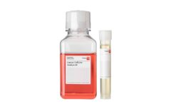
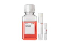
3D Tumorsphere Medium XF
Serum-free and xeno-free medium for the isolation and 3D long-term cultivation of cancer cells.
PromoCell uses the Bioz AI engine to display product citations. This content is currently blocked due to your cookie preferences.
Key benefits:
- Establish 3D tumorsphere cultures directly from 2D cancer cell lines
- Suitable for long-term routine culture of tumorspheres
- Compatible with most commonly used cancer cell lines
- Xeno-free and serum-free formulation
Components:
- 3D Tumorsphere Basal Medium
- 3D Tumorsphere SupplementMix
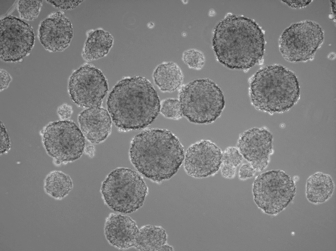
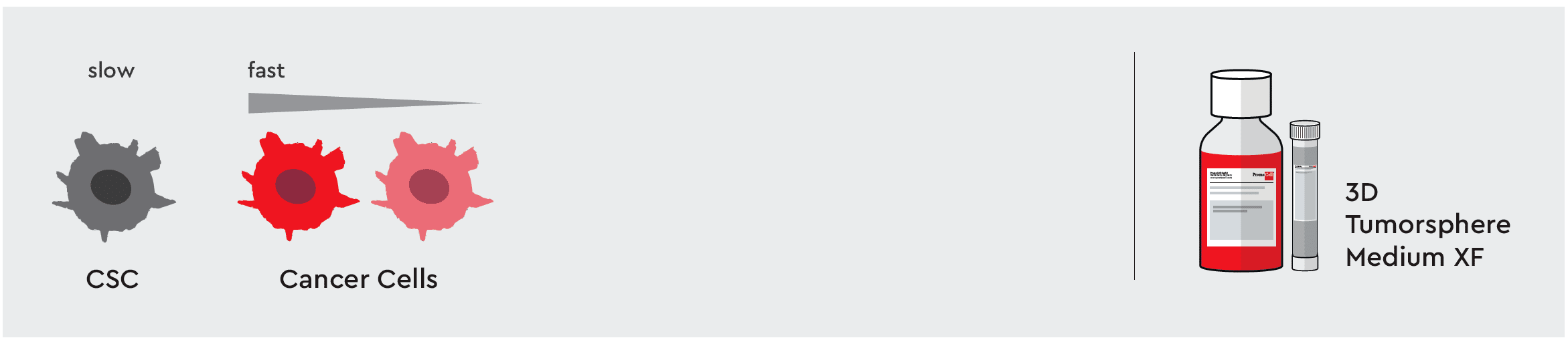
3D cancer spheroids and organoids are popular in vitro models that often better reflect the in vivo tumor environment than some 2D models. Within tumors and tumor spheroids, cancer stem cells (CSCs) are a small subpopulation of cells that play a key role in tumor initiation, recurrence, metastasis, and even resistance to cancer therapies. CSCs exhibit stem cell-like characteristics including self-renewal capabilities and tumorigenicity.
As a part of our Cancer Media Toolbox, our 3D Tumorsphere Medium XF is ideal for isolation and 3D long-term cultivation of CSCs. 3D Tumorsphere Medium XF supports enrichment and maintenance of cancer stem cells as well as further differentiated cancer cells with high cell proliferation rates.
The medium helps you create a reducible in vivo-like system by promoting the formation of homogenous tumorspheres through serial passaging of the 3D culture.
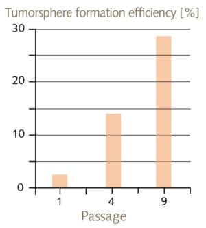
When it comes to the characterization of cultured CSCs, the tumorsphere formation efficiency (TFE) assay represents the gold standard. The TFE assay describes the percentage of cells within a cancer cell culture that can form a sphere from a single cell. Since only stem-like cells can form spheroids from a single cell, TFE values are a quantitative measurement that correlates with cancer metastasis and aggressiveness. In experiment where MCF-7 cells have been cultured in 3D Tumorsphere Medium XF, the TFE value significantly improved from 2% to 28% over 9 passages.
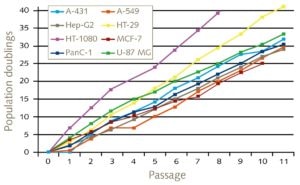
The 3D Tumorsphere Medium XF is flexible enough to support different cell types tested for serial passage:
| Tissue | Tested cell lines | Cell lines origin |
|---|---|---|
| Brain | U-87 MG | Grade IV glioblastoma / astrocytoma of the human brain |
| Breast | MCF-7 | Pleural effusion of metastatic human breast adenocarcinoma |
| Breast | MDA-MB-231 | Pleural effusion of metastatic human breast adenocarcinoma (triple-negative) |
| Colon | HT-29 | Human colon adenocarcinoma |
| Connective tissue | HT1080 | human fibrosarcoma |
| Liver | HepG2 | Hepatocellular carcinoma of the human liver |
| Lung | A-549 | Human lung carcinoma |
| Pancreas | Panc-1 | Epithelioid carcinoma of the human pancreatic duct |
| Prostate | LNCaP | Lymph node metastasis of human prostate adenocarcinoma |
| Skin | A-431 | Epidermoid carcinoma of the human skin |
| Colon | CT26 | Mouse colon cancer cell line |