Myocyte Cell Culture
PromoCell’s myocyte cell culture portfolio features human striated muscle cells isolated from the adult heart and skeletal muscle tissue. Due to their ability to proliferate in vitro, our cardiac myocytes are widely used for assaying cardiotoxicity as well as in other pharmacological studies. Human skeletal muscle cells can be induced to differentiate into myotubes and are commonly used for investigating tissue repair, neuromuscular diseases, and pharmacological testing.
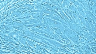
Cardiac myocytes
Cardiac muscle, a unique type of striated muscle in the human heart, is composed of single cardiac myocytes. Because they are responsible for the myogenic contraction of the whole cardiac muscle, cardiac myocytes are the most physically energetic cells in the body. PromoCell’s Human Cardiac Myocytes are produced at from normal human ventricle tissue of the adult heart. Unlike freshly isolated rod-shaped myocytes, PromoCell HCM are isolated using a special protocol allowing them to proliferate in culture over several passages in our Myocyte Growth Medium.
Skeletal muscle cells
New skeletal muscle cells originate from quiescent satellite cells, which reside in the muscle fibers between the basal lamina and the sarcolemma. Quiescent satellite cells are activated by stimuli such as muscle damage. After activation, the cells, now called myoblasts, start to proliferate and fuse with damaged muscle fibers or with one another forming new myotubes. PromoCell’s Human Skeletal Muscle Cells are produced from normal human skeletal muscle tissue from different locations, e.g. M. pectoralis major or M. gluteus maximus (lot specific source information is available on request). The cells can culture over several passages and also be induced to differentiate into multinucleated syncytia using our optimized growth and differentiation media.
Quality control
In addition to cell type-specific marker analysis, each produced lot is tested for growth performance through multiple passages up to 15 population doublings (PD) under culture conditions without antibiotics and antimycotics. In addition, all cells have been tested for the absence of HIV-1, HIV-2, HBV, HCV, HTLV-1, HTLV-2 and microbial contaminants (fungi, bacteria, and mycoplasma).