2")


Human Dermal Lymphatic Endothelial Cells (HDLEC)
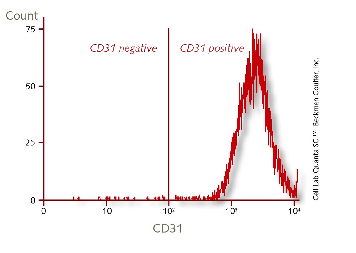
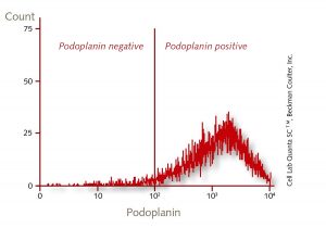
Primary Human Dermal Lymphatic Endothelial Cells isolated from different sources (e.g. juvenile foreskin). CD31 positive, Podoplanin positive.
PromoCell uses the Bioz AI engine to display product citations. This content is currently blocked due to your cookie preferences.
- Product Description
- Additional Information
- Data & Figures
- Technical Library
- Reference Literature
- Downloads
Primary Human Dermal Lymphatic Endothelial Cells (HDLEC) are a subpopulation of the Human Dermal Endothelial Cells. They are isolated from the dermis of juvenile foreskin and adult skin (different locations) from a single donor. The cells are analyzed positive for CD31 and podoplanin by flow cytometric analysis.
(Human Dermal Blood Endothelial Cells (HDBEC) from the same donor are available on request.)
Lymphatic vessels transport excess fluids from tissues to the circulatory system and are a major component of the immune system. The transported fluid is nearly cell free. The vessels are highly permeable, because they lack a continuous basal membrane. Lymphatic endothelial cells are involved in pathological alterations of the lymphatic system. During tumor lymphangiogenesis, the lymphatic endothelial cells build new vessels that infiltrate tumors, attract tumor cells, and induce tumor cell metastasis.
NEW:
- Request our GMP compliant cell culture media for endothelial cells.
- Our HDLEC are now also available from HLA-typed donors.
Available formats:
- Cryopreserved: Cryogenic vial containing 500.000 viable cells.
- Proliferating: >500.000 viable cells shipped in growth medium (T25 flask).
- Cell pellet: 1 million cells dissolved in 200µl RNAlater© for subsequent RNA, DNA or protein analysis. Cell pellets cannot be revived.
| Recommended plating density | 10,000 – 20,000 cells per cm2 |
|---|---|
| Passage After Thawing | P2 |
| Tested Markers | Podoplanin positive, CD31 positive |
| Guaranteed Population Doubling | > 15 |